
3B MICROanatomy Muscle Fiber Model
3B Scientific
- Authorized Distributor
- Fast Delivery
- Easy Returns
MPN :
Muscle Fiber 10,000 Times Magnified - Each - #B60
List Price: $529.18
You Save: $42.33 (8%)
$486.85
Description
Manufacturer Description:
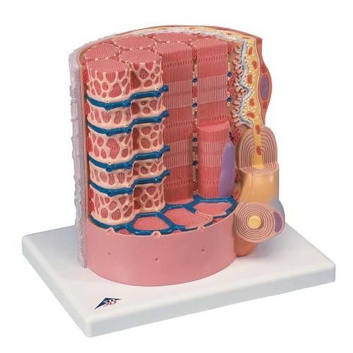
This micro-anatomy model magnifies the anatomy of the human muscle fiber approximately 10,000 times. This muscle model illustrates a section of a skeletal muscle fiber and its neuromuscular end plate. The muscle fiber is the basic element of the diagonally striped skeletal muscle. You've never seen a muscle fiber in this way! This high quality muscle fiber replica brings a hands-on understanding of the human muscle to any classroom. See the anatomy of the human muscle is a whole new way.
Product Videos
Muscle Cell (2:25 Minutes)
Video Transcription
This is a model of a single muscle cell or myofiber. This is just a portion, students often get confused about what on the model is not a fascicle. This would be the endomysium, underneath it would be the sarcolemma which is a plasma membrane of the muscle cell. We have a motor neuron coming in here, it's myelinated we can see the myelin sheath. At the synaptic involved, we see lots of vesicles filled up with acetylcholine, the neurotransmitter. The only neurotransmitter used at the neuromuscular junction and its release through exocytosis which takes energy so you see lots of mitochondria indicating that. As it's released, it diffuses across, there's a little gap here, a synaptic cleft, to the other side. The other side is on the muscle. This is called the motor endplate. They don't show the receptors here but receptors for acetylcholine on this side. When acetylcholine binds, it actually opens up sodium gated channels. Sodium ions rush in because of greater concentration outside than inside. A short time later, there would be opening of potassium gated channels and potassium would rush out. Polarizing the membrane but the sodium rushing in, depolarizing the membrane and that spreads along the surface of the muscle cell. When it gets to the T tubules, it goes deep into the side of the cell and depolarizes the membrane here as well so what we are seeing here is the transverse tubules returning a message deep inside. You noticed here and here is what we called the thermo cisterns of the sarcoplasmic reticulum. So we have calcium ions store here, when the message gets deep inside then the calcium is released. Now, there is an error I think in the information that comes with this, so be careful. This should be the dark band, the A band. This should be the I band. At the junction of the A and I is where you find the T tubules in skeletal muscles. The Z just could be in this area right here.
Additional Information
| Manufacturer | 3B Scientific |
|---|
Login and Registration Form