
3B MICROanatomy Digestive System Model
3B Scientific
- Authorized Distributor
- Fast Delivery
- Easy Returns
MPN :
20-Times Magnified - Each - #K23
List Price: $571.57
You Save: $45.73 (8%)
$525.84
Description
Manufacturer Description:
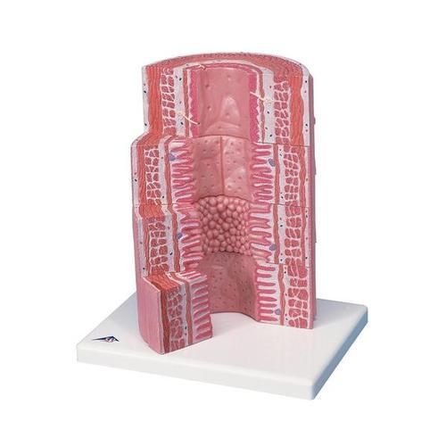
The 3B MICROanatomy Digestive System model illustrates the structure of the fine tissues of four characteristic sections of the digestive system:
- Esophagus
- Stomach
- Small intestine
- Large intestine
The front of the 3B MICROanatomy Digestive System model, from top to bottom, shows a magnified view in histological section of the individual sections of the digestive system and their fine tissue structures. On the back of the digestive system model, highly magnified views of didactically interesting areas of each of the digestive system sections shown on the front are emphasized. The anatomy of the human digestive system is magnified 20 times.
Product Videos
Digestive System Wall Model (3:25 Minutes)
Video Transcription
Let's take a look at this model which is going to show the layering of arteries and veins. If we look at the artery which is this pink structure here in the middle and we got a vein on either side of it. This artery has blood represented with red in the middle that would be flowing in this direction based on the way the valves are oriented in the veins, so blood flowing this direction. In contact with that blood is endothelium and then just outside or superficial to the lumen from there is a subendothelial layer or subendothelium, right here, it's kind of bluish hand color, wrapped around that subendothelial layer is an internal elastic membrane followed by the tunica media or the smooth muscle portion of the artery and then for this artery around the smooth muscle is an external elastic membrane and then finally, the whole artery gets wrapped in adventitia. Veins are slightly different. The artery has no valves anywhere through there but if we take a cross section of the vein, you can see that it does have valves which are going to force blood to flow just in one direction. If any blood was trying to push this way, it would close the cusps on this valve and prevent it from flowing backwards. That's helpful because there's not nearly as much pressure in veins as there are in arteries so the pressure radiant really helps things flow easily in arteries whereas with veins, we rely on skeletal muscle contraction that kind of milk the blood back towards the heart. Layering, veins still have an endothelium and a subendothelial layer. They still have the internal acid membrane and a tunica media but they lack an external elastic membrane. So, you'll notice this layer which is on the artery is not present on the vein. Also noticed, that the veins tunica media, that smooth muscle, is much thinner than it was on the artery. Of course, it still has an adventitia to wrap the whole thing.
Additional Information
| Manufacturer | 3B Scientific |
|---|
Login and Registration Form